Mesothelioma Differential Diagnosis Radiology - Clinical Staging Of Malignant Pleural Mesothelioma Current Perspectiv Lctt / There are other conditions that can mimic pc, such as pseudomyxoma peritonei, peritoneal lymphomatosis, peritoneal malignant mesothelioma, leiomyomatosis peritonealis disseminata, and tuberculous peritonitis.
Mesothelioma Differential Diagnosis Radiology - Clinical Staging Of Malignant Pleural Mesothelioma Current Perspectiv Lctt / There are other conditions that can mimic pc, such as pseudomyxoma peritonei, peritoneal lymphomatosis, peritoneal malignant mesothelioma, leiomyomatosis peritonealis disseminata, and tuberculous peritonitis.
Mesothelioma Differential Diagnosis Radiology - Clinical Staging Of Malignant Pleural Mesothelioma Current Perspectiv Lctt / There are other conditions that can mimic pc, such as pseudomyxoma peritonei, peritoneal lymphomatosis, peritoneal malignant mesothelioma, leiomyomatosis peritonealis disseminata, and tuberculous peritonitis.. The gross findings at the time of surgery or autopsy are extremely useful and these can often be inferred from radiologic studies. Computed tomography is the most commonly used modality for tumor staging. Utility and pitfalls of immunohistochemistry in the differential diagnosis between epithelioid mesothelioma and poorly differentiated lung squamous cell carcinoma. Benign conditions such as scarring or inflammation from prior infections. Benign tumors of the pleura.
The differential diagnosis includes metastatic disease, primary papillary serous carcinoma of the peritoneum, lymphoma, and granulomatous disease. 1 ) and present in 70% of cases 3 , there are two main mechanisms causing ascites: Earlier intervention increases the odds of surviving mesothelioma. Imaging evaluation is essential in diagnosis, staging, and assessment of treatment response in malignant pleural mesothelioma. Ct scan imaging and the diagnosis of mesothelioma when a person with the occupational history of high asbestos exposure and symptoms like relentless coughing, wheezing, chest pain and sudden weight loss, come to see a physician;
Cystic Mesothelioma Of Gallbladder Lodge An Exceptional Location from slidetodoc.com Based on a pictorial review of ct imaging, we will describe its key signs as well as the main differential diagnoses encountered in current practice. Cancers systematic review new updates of the imaging role in diagnosis, staging, and response treatment of malignant pleural mesothelioma chiara romei 1,*, salvatore claudio fanni 2,*, federica volpi 2, alessio milazzo 2, caterina aida d'amore 2, leonardo colligiani 2, emanuele neri 2, annalisa de liperi 1, giulia maria stella 3 and chandra bortolotto 4 citation: Role of ct, mri, and pet/ct in staging evaluation and treatment considerations. Precise diagnosis based on imaging alone is often difficult and very often the final diagnosis is only obtained after appropriate histopathology or microbiology. Pleural mesothelioma is a frequent enough tumor to be considered in the differential diagnosis of chest tumors. The ultrasound can show if the patient has any lumps or masses with 90% accuracy, as well as hydrocele. Benign tumors of the pleura. Malignant mesothelioma should be taken into account in the differential diagnosis of hydrocele, moreover, in patients with a history of scrotal pathology and asbestos exposure.
It may be a symptom of a more severe diagnosis such as malignant pleural mesothelioma.
It is a very rare tumor with nonspecific symptoms (such as chest pain) that can easily be confused with other causes. To distinguish between types, doctors will use imaging scans to examine the patient's lungs and pleura: Although, ct has intrinsic limitations due to low soft tissue contrast and the current staging system as well as criteria for evaluating response, it does not consider the complex growth pattern of this tumor. The etiopathogenesis of bmpm remain unclear. Thus, the differential diagnosis for female pelvic masses is extensive. Main differential diagnosis is metastatic pleuropathy, which have a more "discrete" Benign tumors of the pleura. Tissue sample will be needed to make a definitive diagnosis, although the tumor can spread along the biopsy track. The differential diagnosis of mesenteric lymphadenopathy also includes metastases and. These three ways of identification of symptoms : Cancers systematic review new updates of the imaging role in diagnosis, staging, and response treatment of malignant pleural mesothelioma chiara romei 1,*, salvatore claudio fanni 2,*, federica volpi 2, alessio milazzo 2, caterina aida d'amore 2, leonardo colligiani 2, emanuele neri 2, annalisa de liperi 1, giulia maria stella 3 and chandra bortolotto 4 citation: Scrotal mesothelioma is very rare and less common than pleural or peritoneal involvement; The membrane directly covering the lung tissue
Utility and pitfalls of immunohistochemistry in the differential diagnosis between epithelioid mesothelioma and poorly differentiated lung squamous cell carcinoma. Pericardial mesothelioma is a cancer that begins in the membranes (mesothelium) that surround the heart (the pericardium). The differential diagnosis of mcpm includes other peritoneal cystic lesions, such as pseudomyxoma peritonei, cystic lymphangioma and peritoneal inclusion cyst. Similarly, calcification of the peritoneal thickening makes mpms less likely. Imaging, biomarkers, and the biopsy are also used for cancer in general and not just for epithelioid mesothelioma in particular.
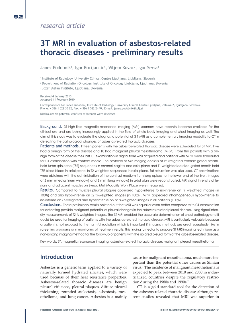
Pdf 3t Mri In Evaluation Of Asbestos Related Thoracic Diseases Preliminary Results from i1.rgstatic.net Multimodality imaging for characterization, classification, and staging of malignant pleural mesothelioma. These diseases may appear similar on computed tomography (ct), but there are some clues for the differential diagnosis. These three ways of identification of symptoms : Ct is the first imaging technique used for diagnosis, staging, and assessment of therapy response. However, there are not specific diagnostic criteria to perform differential diagnosis between metastatic pleuropathy and mpm; It is a very rare tumor with nonspecific symptoms (such as chest pain) that can easily be confused with other causes. It may be a symptom of a more severe diagnosis such as malignant pleural mesothelioma. Based on a pictorial review of ct imaging, we will describe its key signs as well as the main differential diagnoses encountered in current practice.
Precise diagnosis based on imaging alone is often difficult and very often the final diagnosis is only obtained after appropriate histopathology or microbiology.
When confronted with a potential mesothelioma, one needs to know as much clinical and radiologic information as is available. However, the site of origin, mr imaging characteristics, and clinical history may all help narrow the differential diagnosis. Peritoneal mesothelioma prognosis may vary depending on an individual's case, with life expectancy ranging from two to six years. mesothelioma is a neoplasm arising from mesothelial cells that line serous cavities, such as the pleura and peritoneum. mesothelioma occurs in about 2,000 patients a year in the u.s. The diagnosis is also challenging, and often requires a combination of imaging, echocardiogram, and a biopsy. Pericardial mesothelioma is a cancer that begins in the membranes (mesothelium) that surround the heart (the pericardium). Malignant mesothelioma should be taken into account in the differential diagnosis of hydrocele, moreover, in patients with a history of scrotal pathology and asbestos exposure. differential diagnosis may be required for the judgment of compensation, if the pathologic diagnosis is uncertain or the patient is incapable of receiving a biopsy. Exceptionally rare diagnosis localized disease (resembling wdpm) complex papillary or solid architecture, identical to that seen in diffuse malignant peritoneal mesothelioma at least moderate cytologic atypia in most cases increased mitotic activity (> These three ways of identification of symptoms : Pleural effusion has a broad differential as well. Senyigit and colleagues mentioned in their study that although ct findings of mpm vary, they may provide a valuable clue to the diagnosis, at least in patients with a history of asbestos exposure ( 7 ).
In these cases, doctors may perform a testicular mesothelioma ultrasound to observe any irregularities in the testes or scrotum. It is the most accurate test not only for verification of mesothelioma but also in differential diagnosis. Unlike in pleural mesothelioma, associated calcified peritoneal plaques are uncommon. Precise diagnosis based on imaging alone is often difficult and very often the final diagnosis is only obtained after appropriate histopathology or microbiology. There are other conditions that can mimic pc, such as pseudomyxoma peritonei, peritoneal lymphomatosis, peritoneal malignant mesothelioma, leiomyomatosis peritonealis disseminata, and tuberculous peritonitis.
Radiologic Considerations And Standardization Of Malignant Pleural Mesothelioma Imaging Within Clinical Trials Consensus Statement From The Nci Thoracic Malignancy Steering Committee International Association For The Study Of Lung Cancer Mesothelioma from els-jbs-prod-cdn.jbs.elsevierhealth.com Sarcomatoid and biphasic subtypes are less common. Pleural mesothelioma is much more common than peritoneal mesothelioma. The gross findings at the time of surgery or autopsy are extremely useful and these can often be inferred from radiologic studies. Utility and pitfalls of immunohistochemistry in the differential diagnosis between epithelioid mesothelioma and poorly differentiated lung squamous cell carcinoma. The presence of marked and enlarged lymphadenopathy favors lymphoma or metastatic disease as the most likely diagnosis. Imaging evaluation is essential in diagnosis, staging, and assessment of treatment response in malignant pleural mesothelioma. Nickell lt jr et al: 1 ) and present in 70% of cases 3 , there are two main mechanisms causing ascites:
2 department of radiology, dongguk university ilsan.
It is difficult to differentiate mesothelioma from carcinomatosis with imaging alone; Appropriate narrowing of the differential diagnosis is important for optimal patient management. Epithelioid mesothelioma is the most frequent histologic type of malignant mesothelioma; Pleural mesothelioma is much more common than peritoneal mesothelioma. However, absence of findings of asbestos exposure and absence of a mass or metastatic disease elsewhere suggest mesothelioma. While including malignant mesothelioma in his differential diagnosis, the physician go through various tests like ct. The limitations and relative advantages of different imaging techniques are highlighted, and the imaging findings of mesothelioma with each of these techniques are elaborated. Assessment of tumor extension and lymph node involvement is essential in imaging evaluation as locally advanced tumors are amenable to resection. The differential diagnosis of mesenteric lymphadenopathy also includes metastases and. Tunica vaginalis is lined by mesothelial cells; Sarcomatoid and biphasic subtypes are less common. 1 mitosis per 10 high power fields is typical) Pet imaging is now becoming an important part of the diagnosis and evaluation of mesothelioma.

0 comments